CT Scan to 3D Printed Human Heart
3D printing has had a number of impacts on the medical community ranging from anatomical models, to custom implants, to the bio printing of fully functional organs. I’ve been interested in 3D printing for years, and I was thrilled to be able to work with the Mayo Clinic’s 3D Printing and Anatomical Modeling Lab to create a custom model of the human heart. The heart is currently being used for physician education at the Mayo Medical School. This model specifically focuses on the blood within the heart and surrounding vessels so as to highlight the more delicate structures.
Data Acquisition
The raw data for the project was gathered using a Computerized Tomography (CT) scan. Contrast was given via an IV to help highlight the cardiac blood volume. At a high level, CT scanners work by combining a series of X-ray images to create 3D models of internal tissue. This becomes more complicated with the heart, because its continuous heartbeat moves the tissue for each X-ray image taken. Gated CT scans avoid this issue by synching X-ray capture with the beating of the patient’s heart using an ECG monitor.
Data Segmentation
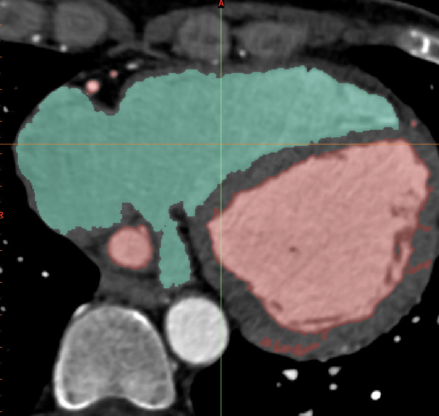
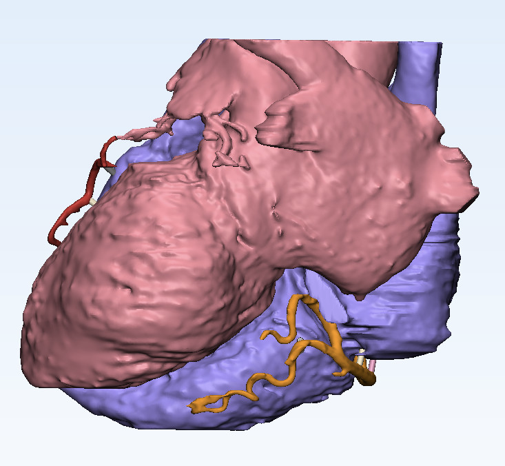
After completing data capture, we moved to segmenting. We leveraged Mimics to select and separate the blood volume into the left and right halves. Red was used to demarcate the left oxygenated side, and blue was used for the right non-oxygenated half.
Support Structure Addition
Following segmentation the 3D model was exported and brought into 3Matics, a Computer Aided-Design (CAD) platform. Using 3Matics, we added additional support structures to the delicate carotid arteries and magnet attachment points.
Printing
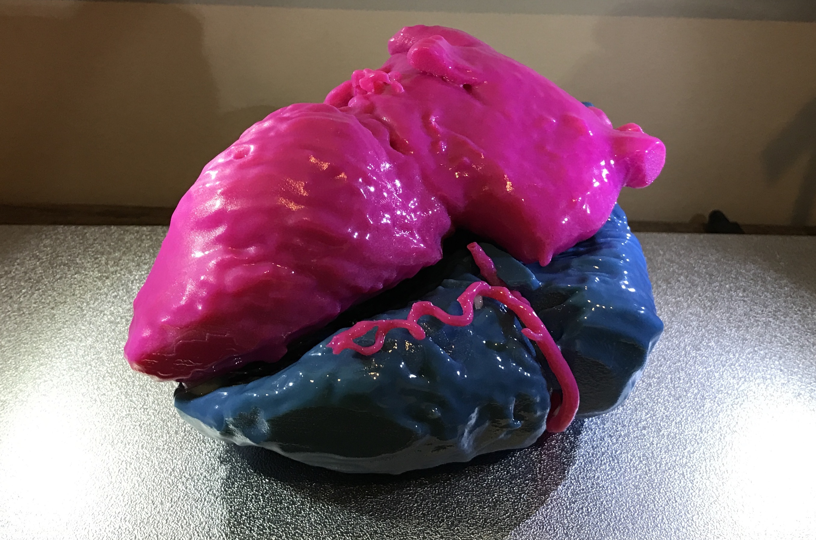
With the model complete, we moved to printing. A Stratasys Objet500 3D printer was used to print the Model. A lye bath was used to remove the extraneous scaffolding material, and the magnets were added to the model.